Upper Leg Tendon Anatomy - Recommendations For Sensor Locations In Hip Or Upper Leg Muscles - All of these tendons protect and house the four ligaments inside of your knee, including your medial collateral ligament, lateral collateral ligament, anterior cruciate ligament and.
Upper Leg Tendon Anatomy - Recommendations For Sensor Locations In Hip Or Upper Leg Muscles - All of these tendons protect and house the four ligaments inside of your knee, including your medial collateral ligament, lateral collateral ligament, anterior cruciate ligament and.. Note that the left gastric artery also gives off an oesophageal branch, which passes through the oesophageal hiatus of the diaphragm to supply the lower oesophagus. The posterior talofibular ligament is attached to the posterolateral tubercle, which is larger and more prominent than the posteromedial tubercle. Percutaneous achilles tendon lengthening is performed in the operating. 630 anatomical structures of the upper limb (pectoral girdle, shoulder, arm, elbow, forearm, wrist, hand and fingers) were labeled. Blood supply to the foot.
The pads of the machine are situated at the achilles tendon. Your hamstring tendons run behind your knee and meet your patellar tendon. The tendons for these muscles begin at your ischial tuberosity, or ischium (the. Upper limb trauma programme injuries. Tendons are fibrous cords attached to muscles and bone.
Muscles Of The Leg And Foot Classic Human Anatomy In Motion The Artist S Guide To The Dynamics Of Figure Drawing from doctorlib.info All of these tendons protect and house the four ligaments inside of your knee, including your medial collateral ligament, lateral collateral ligament, anterior cruciate ligament and. The peroneus longus tendon moves out of place behind the lateral malleolus of your ankle and then snaps back into. Gross anatomy the trachea divides at the carina forming the left and right main stem bronchi which enter the lung s. There are four muscles in the anterior compartment of the leg. Related posts of muscle anatomy upper leg. Superficial veins of upper limb , anatomy : Marc draws and describes the form and location of the upper leg front position. Upper limb trauma programme injuries.
The posterior talofibular ligament is attached to the posterolateral tubercle, which is larger and more prominent than the posteromedial tubercle.
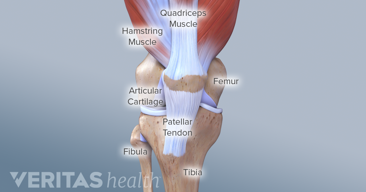
Lateral (fibular) collateral ligament (fcl) upper part middle part lower part popliteus tendon (pt) upper part i. Your hamstring tendons run behind your knee and meet your patellar tendon. Tendon, tissue that attaches a muscle to other body parts, usually bones. Related online courses on physioplus. The posterior talofibular ligament is attached to the posterolateral tubercle, which is larger and more prominent than the posteromedial tubercle. Lie prone on a hamstring curl machine. (from gray's anatomy 40th edition). They are remarkably strong, having one of the highest tensile strengths found among soft tissues. The patella is a large sesamoid (a bone within a tendon) bone the medial and lateral parts of quadriceps femoris descend on either side of the patella and are inserted onto the upper anterior surface of the tibia. It serves to attach the plantaris, gastrocnemius (calf) and soleus muscles to the calcaneus (heel) bone. Tendons are fibrous cords attached to muscles and bone. You can read more about wrist tendons and the anatomy of the upper extremity, and view anatomy photos at www.handcare.org. By spicer mcleroy in tutorials.
Related online courses on physioplus. Note that the left gastric artery also gives off an oesophageal branch, which passes through the oesophageal hiatus of the diaphragm to supply the lower oesophagus. Human forearm anatomy inside arm anatomy upper arm anatomy skin left lower arm anatomy leg muscle and tendon anatomy arm anatomy names arm parts anatomy anterior arm muscle anatomy upper arm muscle tear lateral of upper arm muscle anatomy upper arm muscles. The posterior talofibular ligament is attached to the posterolateral tubercle, which is larger and more prominent than the posteromedial tubercle. Collectively, they act to dorsiflex and invert the foot at the ankle joint.
Guide To Knee Joint Anatomy from embed.widencdn.net Tendons are fibrous cords attached to muscles and bone. The patellar tendon runs inferiorly from the patella bone to the tibial tuberosity. Lie prone on a hamstring curl machine. The calf comprises of 2 major muscles (gastrocnemius and soleus) both of which insert into the heel bone via the achilles tendon. Blood supply to the foot. Anatomy of leg and foot human muscular system stock vector.,category:anatomy of the human leg,muscles of the leg and foot classic human anatomy in motion: Gross anatomy the trachea divides at the carina forming the left and right main stem bronchi which enter the lung s. Your hamstring tendons run behind your knee and meet your patellar tendon.
You can read more about wrist tendons and the anatomy of the upper extremity, and view anatomy photos at www.handcare.org.
The pads of the machine are situated at the achilles tendon. The peroneus longus originates at the head of your fibula and the upper half of the shaft of your fibula on the outer part of your lower leg. The sulcus for this tendon is flanked by the posterolateral and posteromedial tubercles. Tendons are fibrous cords attached to muscles and bone. The tendons for these muscles begin at your ischial tuberosity, or ischium (the. Related online courses on physioplus. 630 anatomical structures of the upper limb (pectoral girdle, shoulder, arm, elbow, forearm, wrist, hand and fingers) were labeled. Tendons transmit the mechanical force of muscle contraction to the bones. Percutaneous achilles tendon lengthening is performed in the operating. The tendons of the edl can be palpated on the dorsal surface of the foot. A tendon is the fibrous tissue that attaches muscle to bone in the human body. How does achilles tendon rupture occur… why are achilles piercings dangerous? Use the mouse scroll wheel to move the images up and down alternatively use the tiny arrows (>>) on both side of the image to move the images.
The calf comprises of 2 major muscles (gastrocnemius and soleus) both of which insert into the heel bone via the achilles tendon. It serves to attach the plantaris, gastrocnemius (calf) and soleus muscles to the calcaneus (heel) bone. Related posts of muscle anatomy upper leg. Lie prone on a hamstring curl machine. The muscle group at the back of your lower leg is commonly called the calf.
Leg Muscles List Anatomy Functions Of Legs Dr Seeds from tailormadehealth.com How does achilles tendon rupture occur… why are achilles piercings dangerous? Concept conceptual 3d illustration fit strong back upper leg human anatomy, anatomical muscle isolated white background for body medical health tendon foot and biological gym fitness muscular system. .16 penile numbness and perineum tenderness.18 any suggested exercises or stretches?.22 leg musculature 209 elbow tendonitis and saddle sores. Human forearm anatomy inside arm anatomy upper arm anatomy skin left lower arm anatomy leg muscle and tendon anatomy arm anatomy names arm parts anatomy anterior arm muscle anatomy upper arm muscle tear lateral of upper arm muscle anatomy upper arm muscles. Upper leg anatomy and function. Upper limb trauma programme injuries. The tendons of the edl can be palpated on the dorsal surface of the foot. Collectively, they act to dorsiflex and invert the foot at the ankle joint.
The tendons of the edl can be palpated on the dorsal surface of the foot.
Percutaneous achilles tendon lengthening is performed in the operating. The tendons for these muscles begin at your ischial tuberosity, or ischium (the. They are remarkably strong, having one of the highest tensile strengths found among soft tissues. This mri wrist coronal cross sectional anatomy tool is absolutely free to use. Fascia of the upper limb. Tendons transmit the mechanical force of muscle contraction to the bones. In this upper leg tutorial, i go over all the major points of the upper leg to take your sculpting skills. Collectively, they act to dorsiflex and invert the foot at the ankle joint. Upper limb trauma programme injuries. This may result in tendon subluxation; The patellar tendon runs inferiorly from the patella bone to the tibial tuberosity. Tendons are fibrous cords attached to muscles and bone. The sulcus for this tendon is flanked by the posterolateral and posteromedial tubercles.


0 Komentar